
Transfrontal craniotomy for surgical removal of frontal meningioma in a canine patient: A case study
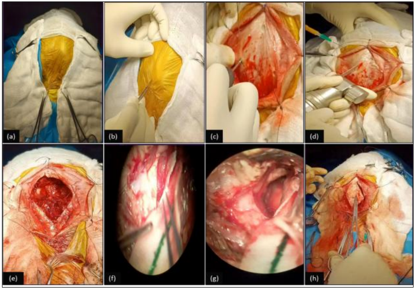
This case report describes the successful surgical management of a frontal meningioma in a 9-year-7-month-old Golden Retriever presented with recurrent seizures. Magnetic resonance imaging and computed tomography identified an extra-axial cystic mass in the left olfactory and frontal region. Preoperative planning incorporated a patient-specific 3D-printed skull model generated from CT-DICOM data. A transfrontal craniotomy enabled near-complete excision of the mass, with histopathology confirming a cystic meningioma, microcystic variant, consistent with WHO Grade I. The patient recovered uneventfully, with complete resolution of seizures and normal neurological status at one-month follow-up. The case highlights the role of advanced imaging, 3D-printed surgical planning, specialized neurosurgical techniques, and comprehensive perioperative management in canine intracranial surgery.