
Design concept of an extracortical implant for canine total hip replacement: A technical note
This technical note presents the Prime Hip Barrel-Plate System, an innovative extracortical, canal-sparing approach to canine total hip replacement. The system uses proximal metaphyseal fixation through a controlled femoral neck tunnel combined with a lateral extracortical plate, preserving the femoral intramedullary canal. The paper describes the implant concept, biomechanical rationale, proposed surgical technique, potential advantages, limitations, failure modes, and future clinical applications. The design aims to reduce stress shielding, eliminate femoral broaching risks, preserve future revision options, and provide proximal load transfer while maintaining the integrity of the femoral canal.
Overview
Canine hip dysplasia (CHD) is a common developmental disorder that can result in progressive degeneration of the coxofemoral joint and severe osteoarthritis. In advanced cases, total hip replacement (THR) can provide a definitive treatment option. However, conventional femoral stem systems occupy the intramedullary canal and may be associated with complications including stress shielding, periprosthetic fracture, stem subsidence, and limitations on future revision procedures.
This technical note presents the Prime Hip Barrel-Plate System, a proposed extracortical and canal-sparing approach to femoral fixation for canine total hip replacement.
Prime Hip System
The Prime Hip system is designed to achieve femoral fixation without broaching or occupying the femoral diaphysis. Instead, fixation is achieved through a proximal metaphyseal bone tunnel combined with a lateral extracortical plate.
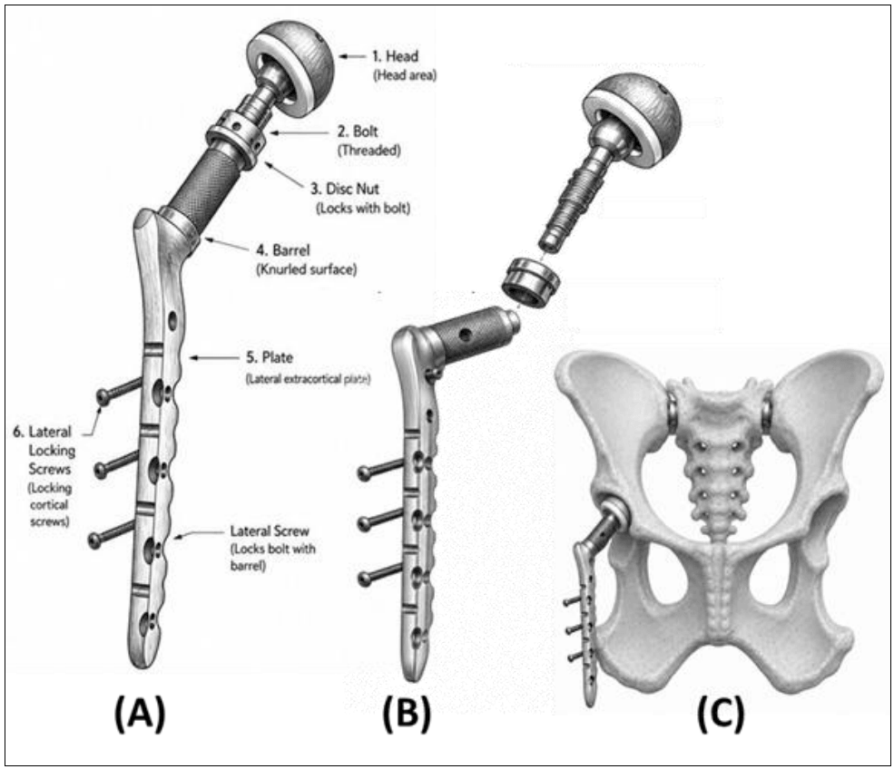
The system consists of three interdependent components: a threaded proximal metaphyseal bolt with disc nut, a lateral extracortical plate with barrel, and the associated femoral head component. The bolt functions as a biological anchor and load-transfer structure, while the plate and barrel provide mechanical stabilization and distribute bending and torsional forces.
Proximal Metaphyseal Bolt
The bolt is positioned through a tunnel created along the natural axis of the femoral neck according to the patient's neck-shaft angle and preoperative planning. The design allows different bolt lengths to accommodate variations in patient anatomy and implant requirements.
The threaded outer surface of the bolt interfaces with a disc nut to distribute load across the proximal femoral bone. The proximal surface accommodates the femoral head component, while the distal portion interfaces with the barrel of the extracortical plate.
Lateral Extracortical Plate
The lateral plate is positioned along the caudolateral surface of the femur and provides extracortical mechanical support. It is designed to help prevent varus collapse, reduce rotational movement at the barrel-bone interface, and distribute bending forces along the outer cortex.
The plate is secured using locking cortical screws after the barrel trajectory has been confirmed. The system is designed to maintain an appropriate three-dimensional trajectory based on the patient's femoral neck-shaft angle and natural femoral anteversion.
Acetabular Component
The acetabular component consists of a hemispherical shell with a textured outer surface designed according to principles used in cementless acetabular fixation. The component is available in different sizes and may incorporate additional screw fixation where press-fit stability is inadequate.
An ultra-high molecular weight polyethylene liner provides the articular bearing surface. The design anticipates osseointegration of the acetabular shell over approximately six to twelve weeks.
Design Rationale
Traditional intramedullary femoral stems transfer load predominantly through the diaphysis and may contribute to proximal stress shielding and bone loss. Femoral broaching can also result in intraoperative fissures or fractures, while stem subsidence and permanent occupation of the medullary canal can complicate future revision procedures.
The Prime Hip system is designed to address these limitations by transferring mechanical forces to the proximal metaphysis and femoral neck while preserving the intramedullary canal.
Biomechanical Principles
The design is based on three fundamental principles: proximal load transfer, preservation of the femoral canal, and extracortical mechanical stabilization.
The extracortical plate distributes bending forces along the outer cortex and provides additional resistance to varus collapse and rotational forces. Preservation of the medullary canal also maintains potential options for future intramedullary fracture fixation or revision arthroplasty.
Safe Cortical Envelope
A critical consideration of the Prime Hip system is preservation of the safe cortical envelope surrounding the planned barrel axis. The femoral neck cortex must remain intact, sufficient cancellous metaphyseal bone must be available to withstand the insertion forces, and drilling must remain axially centered to maintain symmetrical cortical margins.
Oversizing, off-center drilling, incorrect trajectory, or poor bone quality may compromise the integrity of the construct and increase the risk of cortical fissuring or mechanical failure.
Proposed Surgical Technique
Preoperative planning includes calibrated pelvic radiographs and, where available, CT with multiplanar reconstruction. Imaging is used to assess femoral neck geometry, anteversion, cortical thickness, metaphyseal bone quality, and the planned trajectory.
The patient is positioned in lateral recumbency with the operative limb uppermost. A craniolateral approach provides access to the femoral neck, greater trochanter, and proximal lateral femur.
The surgical sequence emphasizes accurate trajectory planning. A pilot hole is created from the lateral femoral cortex along the planned neck axis, with centricity confirmed in both AP and lateral planes. Anteversion is verified against the preoperative plan before progressively enlarging the tunnel.
Once the cortical envelope has been confirmed and the barrel is seated to the planned depth, the extracortical plate is aligned and secured with cortical screws. The bolt is then fixed to the barrel, followed by placement of the disc nut.
The femoral head component is positioned onto the bolt and the femoral assembly is reduced into the acetabular cup. Reduction tension, range of motion, and joint stability are assessed before layered closure. Postoperative radiographs are recommended to verify implant positioning and assess the proximal femur for intraoperative fissures.
Potential Advantages
Femoral Canal Preservation
Because the Prime Hip system avoids occupation of the femoral canal, the diaphysis remains available for potential future procedures. This may be particularly valuable in younger patients who could outlive their initial implant.
Reduced Stress Shielding
Proximal load transfer is intended to preserve more physiological loading of the proximal femoral metaphysis and potentially reduce stress-shielding-associated bone loss.
Elimination of Femoral Broaching
The system replaces conventional femoral broaching with a controlled drill-and-tunnel technique focused on the proximal femur. This may reduce risks associated with intraoperative femoral fissures and fractures during cementless THR.
Applicability to Selected Patients
The design may offer an alternative for patients with poor diaphyseal bone quality, osteopenia, stovepipe femoral morphology, or other anatomical factors that make reliable intramedullary press-fit fixation difficult, provided adequate proximal metaphyseal bone stock is present.
Limitations and Future Directions
The Prime Hip Barrel-Plate System remains in an early stage of development. The authors emphasize that there is currently limited clinical outcome data, cadaveric biomechanical research, and in vivo evidence available for the system.
Potential failure modes are expected to be strongly influenced by implant trajectory and bone quality. An incorrect barrel trajectory may increase loading of the caudal femoral cortex and predispose the neck-calcar region to fissuring or fatigue fracture. Inadequate metaphyseal bone stock may also compromise biological fixation and long-term implant stability.
The technique has a significant learning curve and requires careful three-plane trajectory assessment, surgical simulation, structured training, and appropriate clinical mentorship.
Patient Selection
Potential candidates should have sufficient proximal metaphyseal bone stock, an intact femoral neck cortex, and anatomy suitable for reliable tunnel creation. Severe osteolysis, pathological femoral neck fractures, significantly weakened metaphyseal bone, and extreme patient sizes may make the system less suitable.
Conclusion
The Prime Hip Barrel-Plate System presents a canal-sparing alternative to conventional intramedullary canine total hip replacement systems. Its design addresses several limitations associated with traditional femoral stems, including femoral canal occupation, stress shielding, and broaching-related fracture risk.
The concept relies on precise trajectory planning, preservation of the safe cortical envelope, proximal load transfer, and a strict surgical sequence. While the design provides potential advantages for selected patients, further biomechanical, clinical, and long-term outcome studies are required before its safety and effectiveness can be established.
Publication Information
Authors:Kunal Dev Sharma and Tarun Nar Singh
Journal:International Journal of Veterinary Sciences and Animal Husbandry
Volume:11, Issue 6, 2026
Pages:158-161
DOI:10.22271/veterinary.2026.v11.i6c.3512
Corresponding Author
Kunal Dev Sharma