
Surgical Repair of a Distal Radius–Ulna Fracture in a Canine Patient
A detailed look into a successful treatment at MaxPetZ.
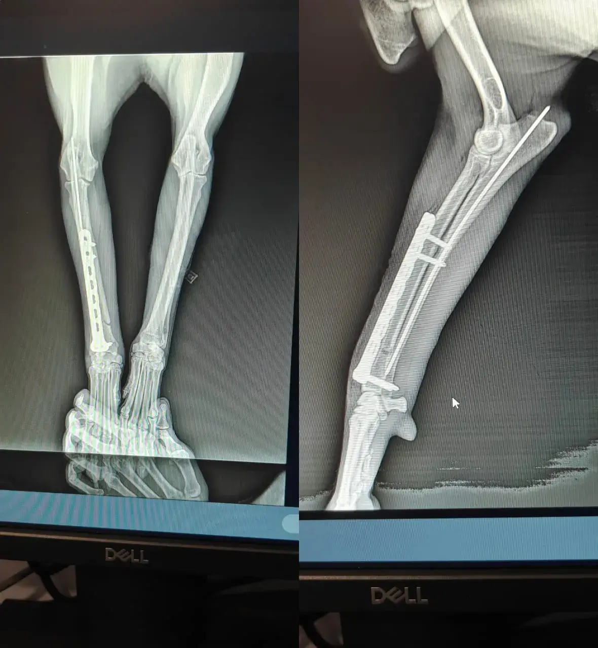
Signalment and History
A 2-year-old, female spayed Labrador weighing 24.2 kg was presented with acute non–weight-bearing lameness of the right thoracic limb after jumping from a sofa. The owner reported immediate crying, holding up the limb, and reluctance to bear weight.
Clinical Examination
The patient was alert and responsive. Moderate swelling and pain were evident over the distal antebrachium. Crepitus was palpable, and the limb was unstable at the distal third. No neurological deficits were noted in the radial, median, or ulnar nerve territories. Cardiothoracic auscultation and abdominal palpation were unremarkable.
"MRI + CT + 3D planning for safer surgery."
Diagnostic Imaging
Orthogonal radiographs of the right radius and ulna revealed a complete, transverse distal-third diaphyseal fracture of the radius with an associated nondisplaced ulna fracture. Mild medial displacement and overriding of the distal fragment were present. The configuration was classified as a simple, complete diaphyseal fracture (Type A3, AO/ASIF classification).
Given the breed and fracture location, conservative management was deemed inappropriate due to high risk of malunion or non-union.
Surgical Procedure
The patient was premedicated with an opioid–benzodiazepine combination, induced with propofol, and maintained on isoflurane anesthesia. A cephalic IV catheter was placed, and perioperative antibiotics (cefazolin) were administered.
A craniomedial surgical approach to the radius was made. The fracture ends were debrided minimally, and anatomical reduction was achieved using fine reduction forceps. Internal fixation was performed with a 3.5 mm locking compression plate (LCP) applied to the cranial surface of the radius. Six cortices were engaged proximally and distally where possible. The ulna was managed with intramedullary pinning at the same time.
Intraoperative radiographs confirmed proper plate position, screw placement, and alignment. The surgical site was lavaged with sterile saline, and routine closure was performed.
Postoperative Management
Analgesia consisted of opioids for 12 hours post-op and NSAIDs for 5 days. A soft padded bandage was applied for 5–7 days to control postoperative swelling. Strict activity restriction was recommended for 6–8 weeks, with controlled leash walking after 3–4 weeks. Jumping and high-impact activities were prohibited.
Outcome
At the 2-week follow-up, the incision had healed normally with no signs of infection. The patient was intermittently weight-bearing with improving limb use.
Radiographs obtained at 6 weeks showed progressing callus formation, maintained alignment, and stable implants. By 8 weeks, the dog demonstrated near-normal limb function with full weight-bearing at a walk.
At the 10-week recheck, radiographic union was achieved. The patient returned to normal activity with no residual lameness.
Discussion
Radius–ulna fractures in dogs commonly occur due to low-energy trauma and have a high incidence of delayed healing or non-union when managed conservatively or when improper fixation methods are used. Rigid stabilization with a plate—preferably a locking system—provides superior biomechanical stability, minimizes complications, and promotes predictable healing. Careful postoperative management is critical due to the thin soft-tissue envelope and limited blood supply in this region.
Conclusion
This case demonstrates successful surgical management of a distal-third radius–ulna fracture in a toy-breed dog using LCP fixation. Early stabilization, precise reduction, meticulous surgical technique, and strict postoperative confinement contributed to an excellent functional outcome.
Treatment Timeline
09 Oct 2025 (admission notes 01:16–01:27)
Initial Presentation / Emergency
Presented with acute onset lameness of right forelimb after play; X-ray confirmed distal radius/ulna fracture. RJ bandage applied; initial emergency stabilization and analgesia given.
09 Oct 2025 (morning)
Diagnostics / Pre-op Testing
CBC, LFT, KFT, Vitamin D3, AFAST and TFAST performed; bloods reviewed prior to anesthesia.
09 Oct 2025 (Surgery performed by Dr. Tarun Nar Singh)
Surgery
Stack T-plate and radius pinning under general anesthesia; routine closure with absorbable deep sutures and non-absorbable skin sutures.
09–10 Oct 2025
Immediate Post-op / Discharge
Post-op medications started (antibiotics, analgesics, supplements). RJ/soft bandage applied; E-collar advised; discharge with home-care instructions and suture removal planned in 10 days.
19 Oct 2025
Suture Removal & Early Follow-up
Suture removal performed. Partial weight-bearing noted. Supplements and medications prescribed (Kalsis, Chymoral Forte, Calcitriol).
08 Nov 2025
Radiographs & Review
X-ray performed (requisition 468926). SOAP: limping on operated limb intermittently; walks normally and bears weight. Pawflex syrup prescribed; plan to review X-ray in 2 weeks and consider implant removal if healing is optimum.
6–10 weeks post-op
Ongoing Recovery
Progressive callus formation and return to near-normal function; final radiographic union by 10 weeks as per standard outcome.
Your Pet Deserves Specialist Care
If you're concerned about your pet's health, our team is ready to provide the advanced care and compassion they need.