
Surgical Management of a Mid-Diaphyseal Humeral Fracture in a Canine Patient
A detailed look into a successful treatment at MaxPetZ.
Signalment and History
A 3-year-old, intact male Labrador Retriever weighing 18 kg was presented with acute non–weight bearing lameness of the left thoracic limb following a vehicular trauma. The owner reported immediate yelping and reluctance to move the limb. No prior orthopedic disease was noted.
Clinical Examination
On physical examination, the patient was bright, alert, and responsive, with normal vital parameters. The left humeral region showed marked pain on palpation, soft tissue swelling, and crepitus. Neurological function of the distal limb (radial, median, and ulnar nerve distribution) was intact. No additional injuries were identified on systemic examination.
"MRI + CT + 3D planning for safer surgery."
Diagnostic Imaging
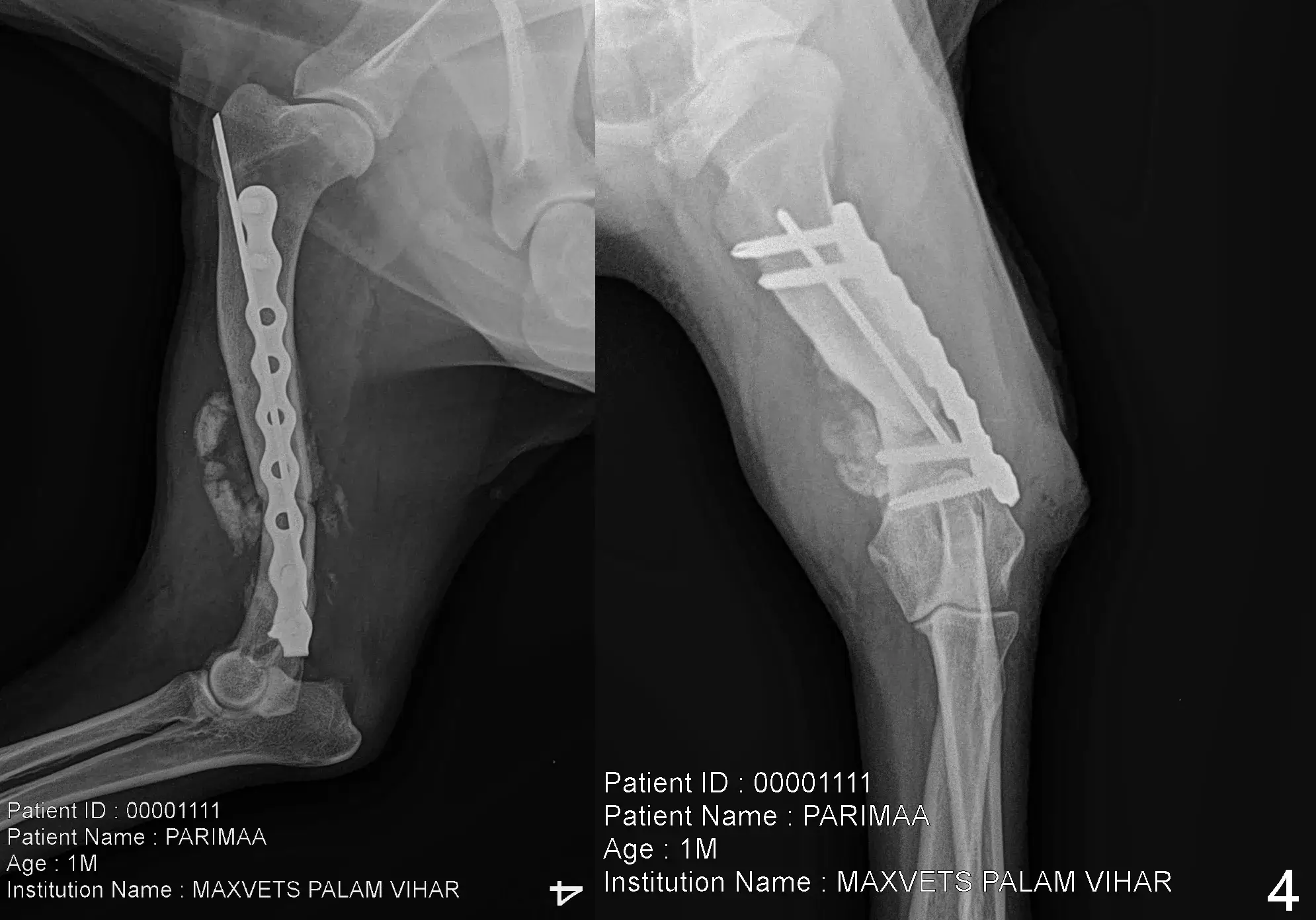
Orthogonal radiographs (mediolateral and craniocaudal views) of the humerus revealed a displaced mid-diaphyseal transverse fracture with mild comminution (two small butterfly fragments). The fracture configuration was amenable to internal fixation.
Surgical Procedure
The patient was premedicated with an opioid–α2 agonist combination, induced with propofol, and maintained on isoflurane anesthesia. Perioperative antibiotics and analgesics were administered.
A standard craniolateral approach to the humerus was performed. Fracture fragments were reduced anatomically using pointed reduction forceps. Internal fixation was achieved using a 3.5 mm locking compression plate (LCP) applied in neutralization mode, with 4 cortices engaged on either side of the fracture. The minor comminuted fragments were preserved in situ as biological scaffolding. Intraoperative fluoroscopy confirmed proper alignment, implant placement, and limb length restoration.
Routine closure of musculature, subcutaneous tissue, and skin was performed. A sterile support bandage was applied.
Postoperative Management
Postoperative analgesia included opioids for 24 hours and NSAIDs for seven days. Strict cage rest was advised for four weeks, with controlled leash walking thereafter. The owner was counseled on preventing premature activity.
Outcome
At the two-week recheck, incision healing was satisfactory with minimal edema. By six weeks, radiographs showed progressive callus formation and excellent implant stability. The patient demonstrated improved weight-bearing and near-normal limb use.
At the ten-week evaluation, radiographic union was achieved, and the dog had returned to full, pain-free mobility.
Discussion
Mid-diaphyseal humeral fractures in dogs are commonly associated with vehicular trauma and often require surgical stabilization because of strong muscle forces causing displacement. Locking plates provide improved stability in diaphyseal fractures, especially when mild comminution is present. Early return to function is typically observed when anatomical reduction and rigid fixation are achieved, combined with appropriate postoperative rest and rehabilitation.
Conclusion
This case highlights successful management of a mid-diaphyseal humeral fracture using LCP fixation. With accurate reduction, stable internal fixation, and carefully monitored postoperative recovery, excellent functional outcome was achieved.
Treatment Timeline
Initial Presentation
Patient presented with non–weight bearing lameness following vehicular trauma.
Diagnostic Imaging
Orthogonal radiographs revealed a displaced mid-diaphyseal transverse humeral fracture with mild comminution.
08 Oct 2025
Surgical Procedure
Humeral fracture repaired using a 3.5 mm locking compression plate (LCP) via craniolateral approach under general anesthesia.
08–09 Oct 2025
Postoperative Care
Analgesia administered; patient monitored; sterile bandage applied.
25 Oct 2025
IPD Boarding
Inpatient care and monitoring continued.
15 Nov 2025
Follow-Up Imaging
Radiographs showed progressive callus formation and implant stability.
Final Recovery
By ten weeks, radiographic union achieved with full, pain-free mobility.
Your Pet Deserves Specialist Care
If you're concerned about your pet's health, our team is ready to provide the advanced care and compassion they need.